Breast Imaging
Breast Imaging
Breast imaging refers to a variety of diagnostic tools used to examine the breasts and detect abnormalities, such as breast cancer, benign tumors, cysts, or infections. The most common breast imaging techniques include mammography (with or without contrast), ultrasound, MRI (Magnetic Resonance Imaging), and in some cases, breast biopsy. Cone beam (CT) of the breast is a new technology which has recently been introduced to Australia.
Purpose of Breast Imaging
Breast imaging is crucial for:
Early detection of breast cancer: Identifying potential issues before they cause symptoms.
Monitoring breast health: For those with a family history of breast cancer or other risk factors.
Evaluating symptoms: Such as lumps, pain, or nipple discharge.
Assessing previous findings: Following up on any suspicious areas or after treatment for breast disease.
Preparing for Breast Imaging
Wear comfortable clothing: You may be asked to remove your top and wear a gown for the procedure.
Avoid deodorants or powders: On the day of your mammogram, avoid applying deodorant, lotion, or powders under your arms or on your breasts, as these products can interfere with imaging results.
Discuss any concerns: Let your doctor or the technician know if you are pregnant, breastfeeding, or have breast implants, as this may affect the type of imaging used or the procedure itself.
Risks and Side Effects
Mammography involves low-dose radiation, but the risk is minimal compared to the benefit of early cancer detection.
Ultrasound and MRI are safe and do not involve radiation.
Biopsy may cause temporary soreness or bruising, but serious complications are rare.
After the Procedure
Mammography/Ultrasound: You can resume normal activities immediately. Results may be available within a few days.
MRI: If contrast dye was used, drink plenty of water to help your body flush out the dye.
Biopsy: You may need to rest for a day, but most patients recover quickly.
Mammography
What is it?
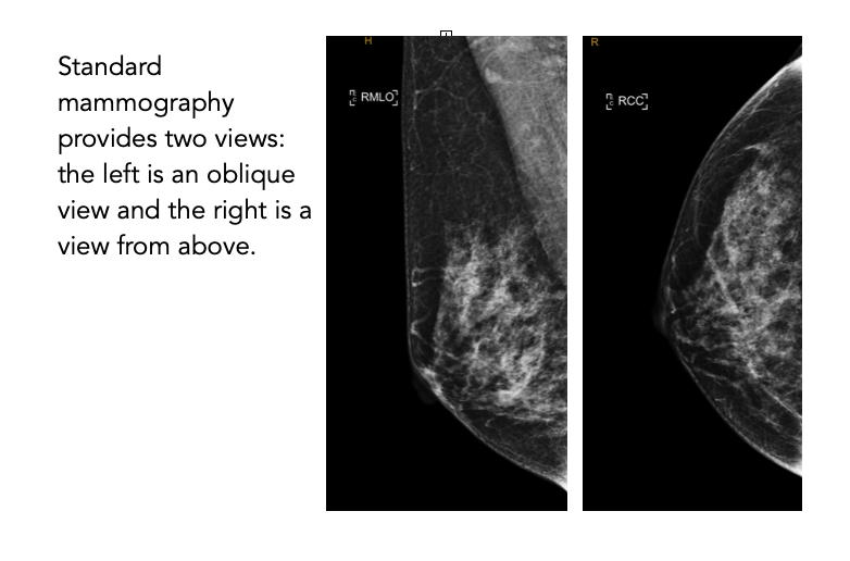
A low-dose X-ray specifically designed to image the breast tissue. It is the most common imaging tool used for routine breast screening.
When is it used?
For regular breast cancer screening, particularly in women aged 40 and older, and to investigate lumps or other symptoms.
What to expect?
During a mammogram, the breast is compressed between two plates to obtain clear X-ray images. The procedure typically takes 15–30 minutes. Some patients may feel temporary discomfort during compression.
Benefits and limitations
Mammography is highly effective for early detection of breast cancer. However, it may be less effective in women with dense breast tissue, which can make it harder to detect abnormalities.
Contrast Mammography
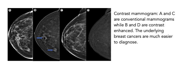
A contrast mammogram is a conventional 3D mammogram (tomosynthesis) which is performed once intravenous contrast is given. The advantage of this technique is the contrast ‘enhances’ any cancerous lesions in the breast, making it much easier to identify. The contrast agent injected into the drip is an iodinated contrast dye (contrast that has iodine). It works by highlighting new blood vessels that form when cancers grow.
There are several scenarios where your doctor may recommend a contrast mammogram:
as a screening test if you have no symptoms but have a higher risk of getting breast cancer. It can also be useful in women who have dense breasts as a screening tool;
to help diagnose breast symptoms;
to gather more information about a tumour you already know you have and to identify if there are any additional hidden tumours.
Cone Beam CT Breast
A cone beam CT of the breast provides a 3D image of the breast. This technique eliminated the issue of overlapping tissue meaning clear and accurate images of the breasts can be obtained. In addition it is pain free with no compression, gives a comparable dose of radiation to standard mammograms, allows whole breast coverage and is safe to use in women with breast implants. A scan of one breast takes seven seconds.
Access to this imaging modality is rare in Australia but we are lucky enough at Brisbane Breast and Surgery Clinic to have access for our patients through Brisbane Radiology.
Cone Beam CT of the Breast
Available at Brisbane Radiology’s Breast Health Imaging Centre
Breast Ultrasound
What is it?
A non-invasive imaging test that uses sound waves to create images of the breast tissue.
When is it used?
Often used as a follow-up to mammography, especially in women with dense breast tissue or to differentiate between solid masses
Breast MRI (Magnetic Resonance Imaging)
What is it?
A powerful imaging technique that uses magnetic fields and radio waves to produce detailed images of the breast.
When is it used?
For high-risk patients (such as those with a genetic predisposition to breast cancer) or to further assess inconclusive findings from mammograms or ultrasounds.
What to expect?
You will lie down inside the MRI machine, which takes multiple images of the breast tissue over 30–60 minutes. Contrast dye may be injected into your veins to enhance the clarity of the images.
Benefits and limitations
MRI is extremely sensitive and can detect even very small abnormalities. However, it is expensive, not suitable for routine screening, and may lead to false-positive findings, requiring further tests.
Breast Biopsy (Imaging-Guided Biopsy)
What is it?
A procedure where a small sample of breast tissue is removed and examined under a microscope. It is often guided by imaging techniques like mammography, ultrasound, or MRI.
When is it used?
When a suspicious area is detected on imaging and needs further examination to determine if it is cancerous.
What to expect?
The doctor will numb the area and use imaging to guide the needle to the abnormal area, from which a small tissue sample is taken. The biopsy itself takes about 15–30 minutes.
Benefits and limitations
This is the only method that can definitively diagnose breast cancer. It is minimally invasive, though some patients may experience minor discomfort or bruising afterward.
Conclusion
Breast imaging is an essential part of maintaining breast health, particularly for the early detection and diagnosis of breast conditions. Your healthcare provider will help you determine which imaging method is best suited for your needs based on your symptoms, age, and medical history.
If you have any questions or concerns, don't hesitate to discuss them with your healthcare team.


